Department of Biological Sciences
Mammalian Histology (B408) is taught in the fall
semester annually and is one of the courses required for the Medical Scholars
Program with the Jefferson Medical College. Consequently, it is taught
at a comprehensive level and concentrates heavily on human tissues and
organ systems. A strong component of this course is tissue structure at
the ultrastructural level and how it relates to structure-functional relationships
at the light microscopic level. A sizeable collection of color light microscopic
images as well as black and white electron microscopic images have been
archived and can be accessed by links with this home page. These images
files have been produced by:
| Dr. Roger C. Wagner
Professor of Biological Sciences
University of Delaware
28499@udel.edu |
 |
| Dr. Fred E. Hossler
Professor of Anatomy and Cell
Biology
East Tennessee State University
College of Medicine |
 |
This page was constructed by
Anuj
Parikh, MSP class of 1998.
In addition, labeled illustrations used in lecture
have been digitized and may be used for study purposes. Three dimensional
models are often useful for understanding the volume structure represented
by two dimensional images and several of these are also linked to this
page.
Click here for COURSE
SYLLABUS and LABORATORY
GUIDE
WAGNER-HOSSLER MICROSCOPIC ANATOMY
IMAGES
Color (8-bit) gif files of light microscopic
images are archived according to tissue type and organ systems. The majority
of these are stained with hematoxylin and eosin but in several cases more
specialized stains are employed. Index pages and files can be accessed
by clicking on red button above.
 Compressed
Histology Images
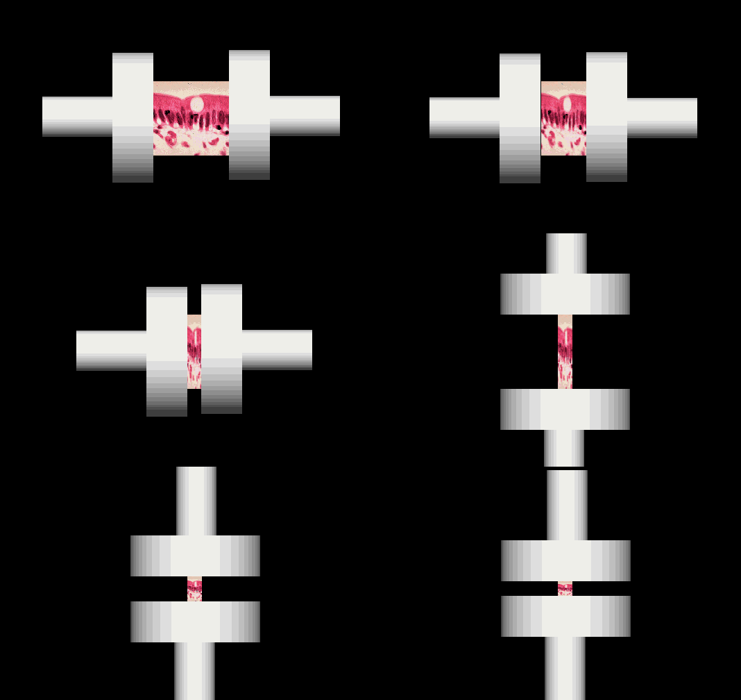
This is an archive of compressed histology images
that can be examined as though the viewer were using a microscope.
These high resolution images have been compressed without loss of image
resolution, resulting in faster downloading times and excellent quality.

 Cell
and Tissue Ultrastructure
Transmission and scanning electron micrographs
are archived as 8 bit grey-scale images and are listed according to tissue
type and organ system. These are meant to compliment the light microscopic
images and reveal structural detail not observable by light microscopy.
Digitized images of overheads of labed illustrations
used in lectures are listed according to lecture schedule and can be broused
for study purposes.
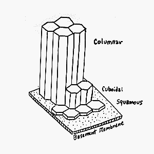
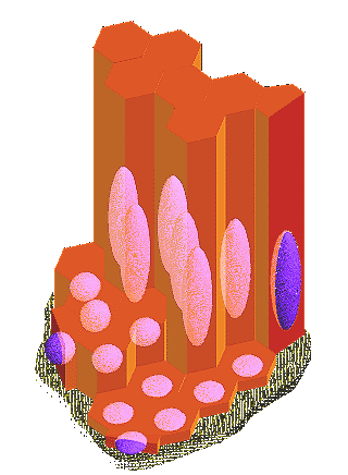
Three dimensional models of tissue and cell structures
have been constructed and rendered to provide realistic interpretations
of volume structures not evident in two dimensional tissue sections.
Links to microscopic anatomy pages
on the WWW:
Histology
World
This page last updated Today.

Compressed Histology Images

